
Videos
- New England Journal
- Sonosite Part 1 and Part 2
1. Vascular Venous Anatomy of Both Arms
 First place a tourniquet above the antecubital fossa to dilate the veins of the lower arm
- Use the linear US probe to identify the cephalic veins over the lateral arm. This is the prefered IV site.
- Follow the veins up and down the arm to find healthy targets
- Look for a large and shallow vein
- Make sure the entire vein is fully compressible to rule out clot
- If no suitable vessels are seen, investigate the veins in the antecubital fossa. Often the median vein can be found in this region
- If no suitable vessels are seen, move the tourniquet close to the axilla, and investigate the veins of the upper arm. Often the cephalic and basilic are good targets
- Veins in order of preference
- Lower arm cephalic
- Lower arm median vein
- Antecubital fossa
- Often has large and superficial veins, but catheters do not last long in the crease of the arm when patients bend their arm frequently.
- Appropriate for short term and urgent use (e.g. GI bleed, severe sepsis)
- Upper arm cephalic vein
- Basilic vein
- Can be found in the lower arm, but difficult to position the patient for successful cannulation
- Often this is the largest vein in the upper arm, and not accompanied by arteries, but can be deep and difficult to access
- Brachial Veins
- These veins are the hardest because they are accompanied by arteries and nerves.
- Should only be accessed by experts


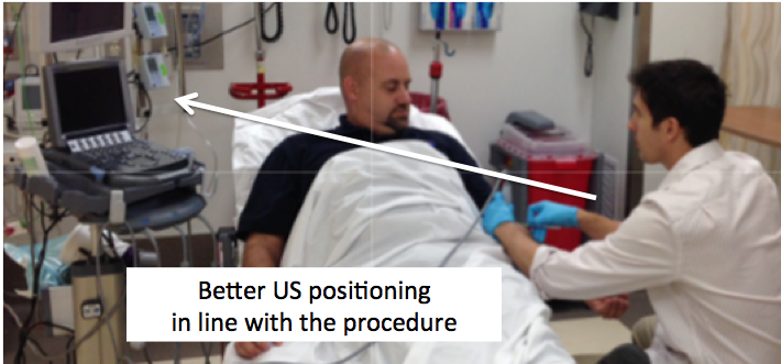
2. Position the patient
- Patient should be supine, and the arm fully extended, 90° to the body.
- Place the ultrasound in line with the procedure, so you can see your hands and the screen without turning your head.
3. Sterile Technique
- US guided catheters last longer than traditional IVs, so additional sterile technique is required per the CDC Guidelines1
- At leasts use sterile gloves, sterile probe cover, and sterile gel.
- Midline catheters
- At minimum use cap/hair net, mask, sterile gloves, a sterile drape, and sterile probe cover and gel
4. Needle Insertion
- Insert the needle at about a 30 degree angle
- More shallow of an angle for superficial vessels < 0.5cm
- Steeper angle for deeper vessels ~ 1cm
- After breaking the skin, find the needle tip on the US before advancing the needle any further. Advance your needle 1-2mm at a time, and always follow the needle tip as it enters the vein.
- Threading the catheter
- Once in the vein, the needle tip will be a clear, crisp bright white dot.
- Hubbing the catheter under ultrasound guidance will increase success rates
- Under ultrasound guidance, advance the needle tip, until it approaches the back wall, then flatten the angle to bring the needle tip back to the center of the vein
- Repeat this advance and flattening technique until the catheter is hubbed or until your are unable to keep the needle safely away from the vessel walls
- Once in the vein, the needle tip will be a clear, crisp bright white dot.
- Gently place the US down, and cast off catheter with 2 fingers of your free hand
- Once the catheter is in, ensure the blood flow is dark, and nonpulsatile. Then apply your sterile finger over the hub to decrease bleeding from IV
- Release the tourniquet
- Attach the catheter extension tubing, aspirate blood, then flush the line with saline
- Saline should flow easily without resistance or pain. If there is not good forward flow, the IV is likely extraluminal or the vessel has been too diseased and damaged to be used.
- Use ultrasound to examine the catheter while flushing if there is concern that IV is not functioning properly
- Use the Tegaderm IV Securement Dressing
- This better secures the IV to skin like a PICC line, preventing dislodgement or bacterial contamination. This allows the catheter to function longer
5. Vascular Access Care1
- Every 7 days, change the dressing every with sterile gloves, and gently cleanse the site with chlorhexidine
- Inspect dressing regularly, and replace dressing if it becomes damp, loose, or is visibly soiled
- Change dressing with sterile gloves, and clean site with chlorhexidine
- Do not routine replace peripheral IV unless there is sign of phlebitis or they do not flush well.1,2
Reference
- O’Grady NP, Alexander M, Dellinger EP, et al. Guidelines for the prevention of intravascular catheter–related infections. Clinical infectious diseases 2002;35:1281-307.
- Rickard CM, Webster J, Wallis MC, et al. Routine versus clinically indicated replacement of peripheral intravenous catheters: a randomised controlled equivalence trial. The Lancet 2012;380:1066-74
- Introduction to Bed Side Ultrasound: Volumes 1 and 2. M Dawson, M Millan. Emergency Ultrasound Solutions. 2012.
- Ultrasonography Assisted Peripheral Line Placement. Jehangir M Mee. Gowthaman Gunabushanam. http://emedicine.medscape.com/article/1433943-overview#a1
